Back to Home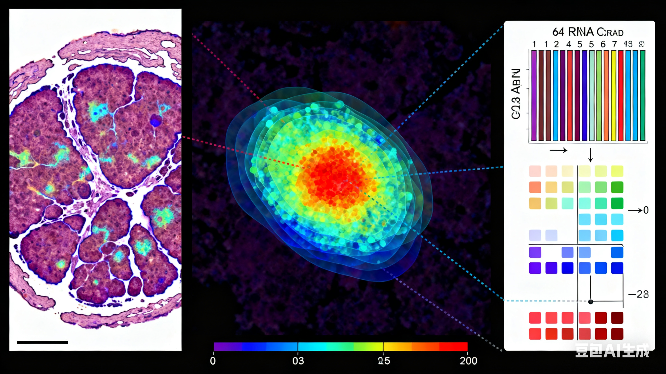


High-plex spatial RNA imaging in one round with conventional microscopes using color-intensity barcodes
By Tianyi Chang, Shihui Zhao, Yanyi Huang•October 30, 2025•
9 min read
•8,150 views
Scientists have developed a groundbreaking technique for high-plex spatial RNA imaging that enables simultaneous visualization of 64 different RNA targets in a single imaging cycle using conventional microscopes. This innovation, based on color-intensity barcoding, dramatically increases the throughput and accessibility of spatial transcriptomics.
Traditional spatial RNA imaging methods are typically limited in the number of targets that can be simultaneously detected, often requiring multiple imaging cycles that increase experimental time and complexity. The new approach developed by Tianyi Chang, Shihui Zhao, and Yanyi Huang overcomes these limitations by using combinations of different fluorophores at varying intensities to create a barcode system that can uniquely identify dozens of RNA targets in a single experiment.
This advancement has profound implications for biomedical research, enabling researchers to map the spatial organization of gene expression with unprecedented detail and efficiency. The technique is particularly valuable for studying complex biological systems such as developing tissues, neural circuits, and tumor microenvironments, where understanding the spatial relationships between different cell types and their gene expression profiles is crucial.
Published in Nature Biotechnology on October 30, 2025, this method democratizes high-plex spatial transcriptomics by making it accessible to laboratories with standard fluorescence microscopy equipment.
You May Also Like
View AllSpotify to raise US prices in first quarter of next year, report says
2 minNov 25
OpenAI learned the hard way that Cameo trademarked the word ‘cameo’
2 minNov 24
Speechify adds voice typing and voice assistant to its Chrome extension
4 minNov 25
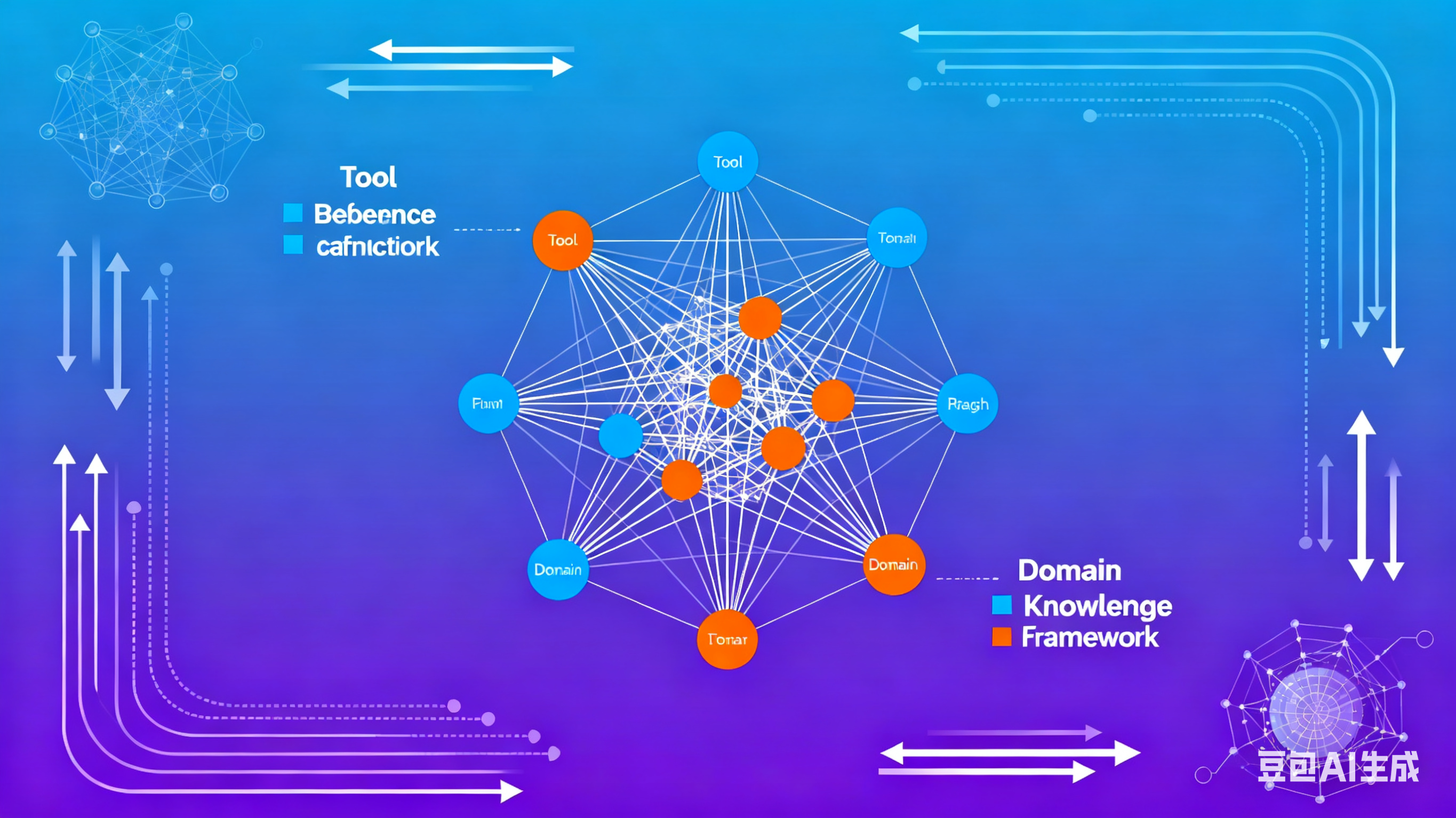
Bridging Tool Dependencies and Domain Knowledge: A Graph-Based Framework for In-Context Planning
12 minOct 28